Damage Assessment

It was December 2015, and Gandy, a neurologist at Mount Sinai Hospital in New York, was showing a former National Football League player named Sean Morey scans of his brain. A professional athlete for 10 seasons, Morey retired from the NFL in 2010 after doctors told him he had suffered too many concussions.
Morey subsequently became a behind-the-scenes health and safety advocate, co-chairing an NFL Players Association committee devoted to brain injuries and leading a mid-2010s effort to improve the settlement terms of a class action concussion lawsuit brought by retirees against the league. Just 39 years old, he also was suffering debilitating headaches, memory lapses, angry outbursts and other symptoms associated with chronic traumatic encephalopathy (CTE), a neurodegenerative disease linked to repeated blows to the head.
While CTE had been found in the brains of dozens of former players, including Pro Football Hall of Fame members Mike Webster and Junior Seau, there was no way to know if Morey had it. The disease could be diagnosed only posthumously.
Gandy was working to change that. To try to identify CTE, he was using an experimental technique to scan the brains of retired football players and soldiers. The scans were color-coded. Healthy people’s brains appeared mostly blue and green, like grassy islands surrounded by sea. By contrast, probable areas of damage looked yellow and red, as if fires were burning on those same islands. The images Gandy was showing Morey were the results of brain scans done seven months earlier using a positron-emission tomography (PET) machine. They were unlike any brain images Gandy had seen before. (He would go on to publish his case study of Morey, one of the first of its kind, in a scientific journal in 2016.)
Much of Morey’s brain was green. But some areas were red. Bright, flaming red. And those areas corresponded to the damaged areas found in the autopsied brains of Seau, Webster and others with CTE. “I knew they would find something,” Morey told me later. “The question was, what was the extent of it?”
Gandy strongly suspected CTE. Yet he couldn’t say for sure and cautioned Morey not to draw sweeping conclusions. The pictures were merely snapshots, produced by a new and unverified method of peeking through the skull to spot a very specific thing. They didn’t indicate that Morey indisputably had the disease, or that his condition would worsen over time. To know anything with certainty, Gandy explained, many more people would need to be scanned. And much more work would need to be done. Morey’s images were not the last word; instead, they were an important step toward solving a medical puzzle. Namely, can CTE be diagnosed in living people?
Now a 44-year-old assistant high school football coach living in Princeton, N.J., and still coping with the damage done by his professional career, Morey knows better than anyone what an answer could mean — both for a sport grappling with the disease and for the lives of the people who play it.
 |
| Neurologist Sam Gandy at Mount Sinai Hospital in New York in January. Photo by Chris Sorenson. |
Suppose you’re a football player. Here’s what happens when doctors suspect that you’ve torn a knee ligament: They use a magnetic resonance imaging (MRI) machine to look at the tissues inside your leg, determine if and how they’ve been damaged, and use that information to prescribe a course of treatment and recovery. By contrast, here’s what happens when doctors suspect you’re suffering from CTE: They wait for you to die. After that, they carefully remove your brain from your skull, slice certain portions of it into small, thin pieces, stain those pieces with special chemicals and then place them under microscopes to look for the disease’s biological signature: tangles of a toxic, abnormal variation of a protein called tau in the depths of the sulci, which are the crevasses between the brain’s many wrinkles. Then, and only then, can it be said conclusively that you had CTE — and not some other neurodegenerative disease or type of brain injury.
This is a problem. Like society at large, football is now coping with the coronavirus pandemic. But the sport has been gripped by an ongoing health crisis since 2005, when a neuropathologist named Bennet Omalu published a study identifying what he believed to be CTE in Webster’s brain. His discovery inspired the 2015 Will Smith film “Concussion.” A 2017 Boston University study of the brains of 202 deceased former football players found that 110 of the 111 who had played in the NFL had the disease, as did lower percentages of athletes who stopped playing in college (48 of 53) and high school (3 of 14).
Linked to aggression, depression, suicidal thoughts, impaired judgment, impulse control problems and dementia, CTE was found in the brain of Tyler Hilinski, a 21-year-old Washington State University quarterback who died of suicide in 2018, and in the brain of Aaron Hernandez, a 27-year-old former NFL player who in 2017 hanged himself in prison while serving a life sentence for murder. The disease sparked thousands of individual lawsuits that led to the NFL’s settlement, which already has awarded nearly $790 million to retirees with cognitive impairment or conditions such as Parkinson’s or Alzheimer’s. CTE was a major factor in a similar settlement between former college athletes and the NCAA and is a key part of a second wave of football-related suits against the association and various athletic conferences and schools.
Concerns about CTE also have contributed to a nationwide decline in high school and youth tackle football participation, local and state-level efforts to ban both activities, and a number of NFL and college players walking away from the game. “The impact of CTE has been dramatic,” says former San Francisco 49ers linebacker Chris Borland, who in 2015 retired from the NFL at age 24 following his rookie season because of concerns about the disease. “We’re seeing players take it into consideration more and more. And it calls into question a lot of the circumstances surrounding football: How much public money should be contributed to the game, whether or not college athletes should be compensated for doing a risky job, if it’s an appropriate activity for young children.”
Scientists believe that repetitive brain trauma — not just concussions, but also less severe subconcussive blows like the hits football linemen absorb on every snap — is a precondition for CTE. Last year, Boston University researchers found that for football players, both the risk of developing the disease and its severity increase with the number of years playing the sport; athletes whose youth-to-pro careers lasted more than 14.5 years were 10 times as likely to have CTE as those who played fewer.
Yet because there’s no way to identify the disease in the living, many basic and important questions remain unanswered. Researchers don’t know why some people who suffer repeated head hits develop CTE while others do not. They don’t know how many hits are too many. They don’t know exactly how the disease first arises, or how and why it spreads across the brain over time. They don’t know why individuals develop different symptoms with different levels of severity. They don’t know how common CTE is, nor how risky football and other contact sports truly are. Most of the brains given to Boston University and other research institutions for study come from donors and families who were experiencing problems during life, which creates selection bias in studies and means headline numbers like 110 of 111 NFL brains skew high.
Moreover, the inability to detect the disease in people such as Morey makes developing effective therapies almost impossible. But with detection, “We’d be able to begin clinical trials for new compounds to be able to treat the disease once it starts — and hopefully even prevent it if we can detect it early on,” says Robert Stern, the director of clinical research for Boston University’s CTE Center and an expert on the disease. “So the next critical step is to diagnose it during life.”
Gandy joined the quest to find a way to detect CTE in the living after studying Alzheimer’s for decades. He helped to discover the first drugs that reduce the formation of beta-amyloid, a sticky protein that builds up in the brains of Alzheimer’s patients. His interest in brain disease was intellectual, rooted in a desire to go from “no understanding of a situation” to “that sudden flash of insight.”
It also was personal. One of Gandy’s earliest memories is of riding in his parents’ car, following a police cruiser that was taking his grandmother to a South Carolina hospital to be institutionalized for dementia. She would confuse her grandson with his father. She would leave her single-story brick cabin and walk down the highway until police picked her up. Before her death in the late 1960s, she spent her final years largely confined to a rocking chair, singing hymns to herself. “I knew something was going on, clearly stressful and important, but I didn’t really understand,” Gandy says.
Gandy, 63, now believes that his grandmother had Alzheimer’s, which wasn’t considered to be a major disease until the 1970s. Gandy’s mother later was diagnosed with the disease. Before her death in 2018, she became paranoid and absent-minded; nurses often found her on the floor between her wheelchair and bathroom after she would call for assistance, forget that help was on the way, and then unsuccessfully try to reach the toilet herself. “I’m sure that all had an effect on me,” Gandy says. “At some level, [brain disease] almost never leaves my mind.”
In the 1990s, Gandy was part of a research team that discovered professional boxers carrying a particular gene known to increase Alzheimer’s risk were more likely to suffer long-term brain damage from prizefighting than their peers. Over the next decade, he closely followed the emerging connection between football and CTE, in part because one of the researchers who helped find the disease in Webster’s brain, Steve DeKosky, was a longtime friend and colleague.
In 2014, Dave Herman, a 73-year-old retired NFL player with memory problems, wanted to join a Mount Sinai clinical trial for an Alzheimer’s drug. Problem was, the five doctors who examined Herman couldn’t agree on a diagnosis. Based on his symptoms, three believed he had the disease. Two suspected CTE. Gandy, who later published a paper on Herman’s case, scanned Herman’s brain twice with a PET machine, which detects radiation. For the first scan, Herman was injected with a radioactive chemical, called a tracer, that binds to beta-amyloid, the protein involved in Alzheimer’s. For the second scan, Herman was given a tracer that binds to the tau protein. In healthy brain cells, tau acts like a kind of scaffolding and is essential to normal functioning; but in CTE, an abnormal variation called p-tau clumps in the valleys of the brain’s wrinkles, killing cells and disrupting function.
Herman’s first scan came back negative: greens and blues, indicating minimal beta-amyloid concentration. Alzheimer’s was ruled out. By contrast, the second scan showed yellow and red. The tracer was sticking to p-tau. Gandy thought it was likely Herman had CTE, but the quality of the scans made it hard to tell whether the p-tau was concentrated in the same brain valleys as seen in autopsies of people who had the disease. To reach a more definitive conclusion, Gandy would need better scans. And he would need more subjects.
 |
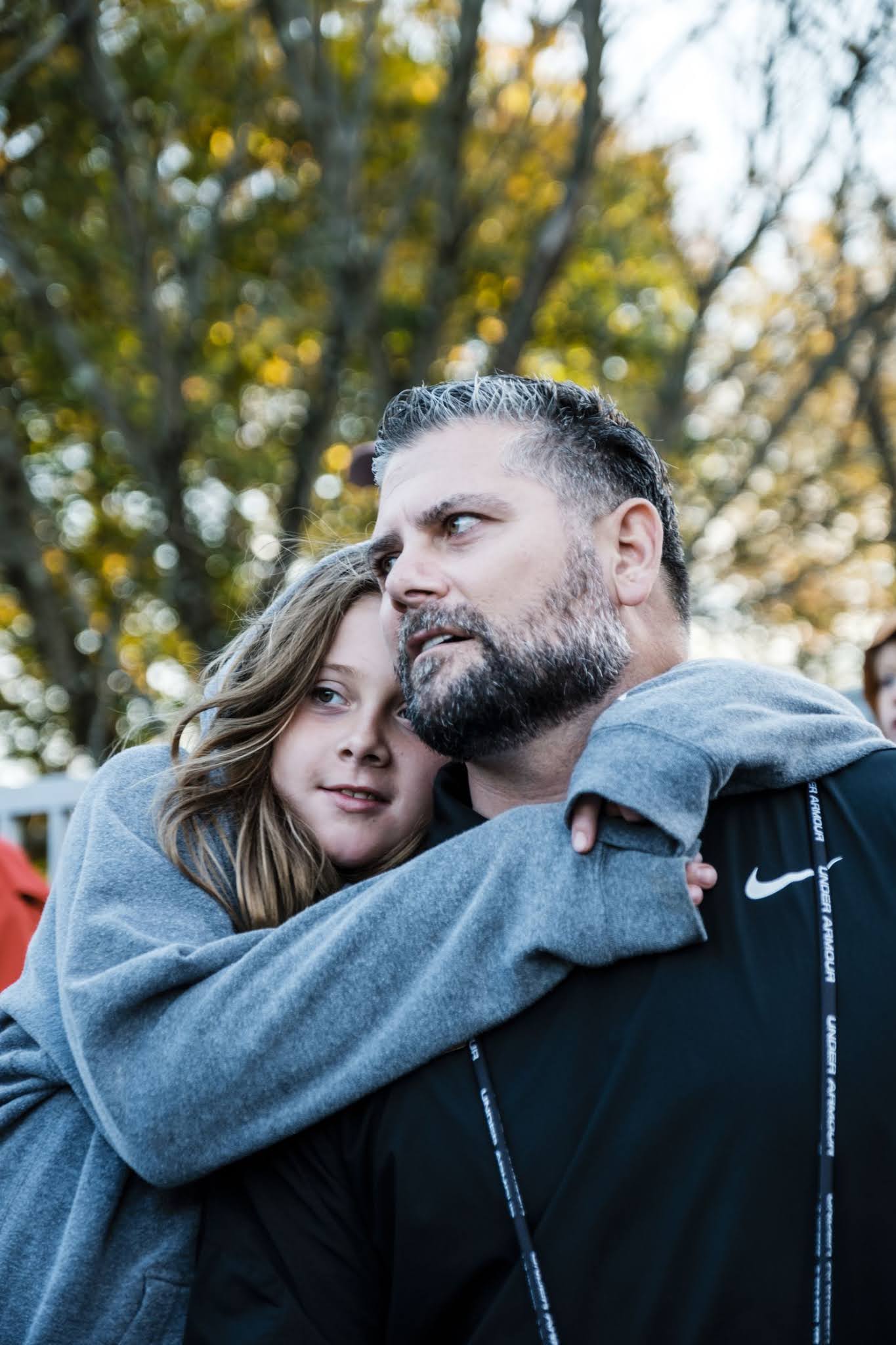
| Morey with daughter Piper after her hockey game in Princeton in November. Photo by Chris Sorenson. |
Morey never planned on becoming a medical case study. Undersized at 5-foot-11 and 193 pounds, he played his way from practice squads to the 2008 Pro Bowl as a hard-hitting special teams standout for the Arizona Cardinals. After football, Morey figured, he would put his Brown University degree to use and become an athletic director. Or perhaps go into politics like Steve Largent, a former NFL receiver who served in Congress. “I wanted to do something positive and serve other people,” Morey says. “Make sure they are not forgotten or misrepresented.”
Brain injuries derailed those ambitions. Morey estimates that he suffered more than 20 concussions over his career, most of them undiagnosed, and countless other blows to the head. Following the 2009 season, he began having blind spots in his vision and excruciating headaches that would leave him immobilized for hours at a time. “I played in the NFL with ripped-off toenails, dislocated fingers, separated shoulders, broken ribs, patellar tendinitis, torn ligaments in my ankle, a torn biceps,” he says. “But I had never experienced that kind of pain.”
When two doctors advised Morey to retire in the summer of 2010, he knew enough to take their advice. Two years earlier at the Super Bowl, he had had a long conversation with Chris Nowinski, a former Harvard University football player and professional wrestler who was trying to warn the sports world about the potential dangers of concussions and repetitive brain trauma, including CTE. First identified in 1928, the disease had been studied in boxers for decades. But football’s medical community largely hadn’t paid attention. “When Chris and I first spoke,” Morey says, “I couldn’t even pronounce ‘chronic traumatic encephalopathy.’ ”
Morey was a quick study. He co-chaired the NFL Players Association’s first brain injury committee and recruited scientists to advise it. He read every study he could get his hands on and co-wrote a 90-page report outlining health risks faced by players. During 2011 collective bargaining negotiations between the league and the union, he helped create a return-to-play protocol for concussed players and helped secure more than $300 million in new player health care and research funding. (Morey later resigned from the union committee because he felt that the programs the union chose to spend that money on did not adequately study or treat retirees with brain injuries. The NFLPA, which is funding a large and ongoing medical study of former players through Harvard University that includes brain injury research, declined to comment on Morey’s resignation.)
All the while, Morey’s own symptoms were intensifying. His headaches, he says, felt like “being beaten in the head with a rubber mallet. Like someone is squeezing your eyeballs together. Every time the blood pumps through your skull, you literally feel it.” He would lose his train of thought mid-conversation, miss appointments, struggle to sleep and find words, leave the stove on, drive away from gas stations with the refueling nozzle still in his sport-utility vehicle. At his daughters’ sports practices, Morey says, he would “show up and introduce myself and ask [other parents] their names, and they would look at me like I’m crazy — because I take them to practice every week, and I’m asking every week.”
Worse still were the explosions of rage. In locker rooms, Morey took pride in being calm and thoughtful. Now he was erupting at his wife, Cara, and their three young daughters, “dropping f-bombs and screaming at the top of my lungs,” he says. Anything could trigger an outburst, like his children arguing in the back seat during a drive to their grandparents’ house. The fallout left Morey confused and ashamed. “I would think to myself, ‘What the f--- did I just say?’ ” he says. “ ‘How can I do this?’ ”
Five years ago, when Gandy first saw the images of Morey’s brain scans, a single thought went through his mind. That’s it. That’s CTE. The scans were negative for beta-amyloid and positive for p-tau; more important, they were at a higher resolution than Dave Herman’s scans, which meant Gandy could see more clearly that the p-tau in Morey’s brain was concentrated in the areas associated with the disease.
“I was amazed,” Gandy says. Ensuing research has produced a more complicated picture, indicating that a CTE test will take a while to develop. To wit: Since 2016, Gandy and his team have scanned and examined 25 combat veterans and 25 former collision-sport athletes. Of the 20 patients suffering clinical symptoms associated with CTE, 16 have scans showing excess retention of the tracer that indicates tau tangles.
However, the other four do not — perhaps because they don’t have the disease, Gandy says, or perhaps because they have CTE but haven’t yet accumulated enough p-tau in their brains to register on scans. “The positives were in people who had obvious symptoms, who already had seen a neurologist or a psychiatrist and were being medicated,” he says. “So, is this technique sensitive enough to see if you have a lower level of [p-tau] or early CTE? Would it be useful in screening NCAA [football] players? Probably unlikely.”
A 2019 Boston University scanning study that compared 26 former NFL players suffering from symptoms associated with CTE and 31 people with no history of head injuries or symptoms was similarly suggestive — and similarly inconclusive. At the group level, the football retirees had higher p-tau levels in their brains than the non-players; among individuals, retirees who had played the longest had the highest levels. However, some of the retirees had the same levels of p-tau as individual non-players, and researchers found no correlation between p-tau levels and symptom severity.
Stern, the Boston University researcher and one of the study’s co-authors, believes that the tracer used to detect p-tau, a compound called flortaucipir, may prove inadequate for a reliable CTE test. “It can bind to stuff in and around the brain that is not abnormal tau,” he says, “and that may alter how the scans are interpreted.” Improved compounds are being developed; last year, British scientists found that the p-tau in CTE has a unique atomic structure compared with the p-tau that can build up in other neurodegenerative diseases, including Alzheimer’s, a discovery that they believe could lead to a CTE-specific tracer.
Even if that happens, doctors still will need to scan a large number of suspected CTE patients, track them over time and examine them after death to validate that the yellow and red areas seen in scans correspond to what’s actually inside their brains. That process will likely take years. When researchers previously worked to validate the PET scans now used to detect beta-amyloid in the brains of Alzheimer’s patients, Gandy says, they were able to move relatively quickly because it is easier for doctors to find patients with Alzheimer’s near the end of their lives. By contrast, he says, there are “fewer people with CTE. And the disease isn’t necessarily going to kill them within the next six months.”
But Stern feels a sense of urgency and wants to speed things up. He is heading the largest and most thorough study designed to identify CTE in living people. The seven-year, $16 million project funded by the National Institutes of Health has put 180 former NFL and college football players and 60 noncontact-sport control subjects through a series of tests including two PET scans; two other types of imaging; blood, saliva and spinal-fluid collection; genetic evaluations; neuropsychological testing; and clinical examinations and histories.
One of the study’s goals is to use imaging alongside the other biomarkers to create a detection tool kit for CTE — if a scan alone isn’t enough to diagnose the disease, then perhaps a scan plus, say, high levels of a chemical in the blood that indicates brain trauma will be. Researchers also hope to find a simple, inexpensive screening method to help doctors determine which patients need costly PET scans and other diagnostic procedures, the way doctors looking for prostate cancer use blood tests to decide who needs more thorough examination. “There are great stretches of the world where you can’t plug in a PET scanner,” Gandy says. “Having a blood test [to screen for CTE] would make it more available and convenient.”
Initial testing in Stern’s study was completed in late February, just days before the coronavirus pandemic shut down college campuses and research labs across the country. Working from home and conferring regularly via video chat, researchers in the study are analyzing the data that they’ve collected and expect to submit the first papers reporting their findings to medical journals before the end of the year. “It’s a long road,” Stern says. “The more neuroscience moves forward making discoveries about the brain, the more we realize how complex it is.”
 |
| Gandy in his office lab at Mount Sinai in January. Photo by Chris Sorenson. |
On a cool and sunny Saturday afternoon last fall, Morey stood on the sideline at the Lawrenceville School in Lawrenceville, N.J., holding a kickoff coverage play sheet he designed himself. Since 2016, he has been an assistant football coach at the private prep school, which has been playing the sport since the 1800s. Outside the Lawrenceville locker room, players lined up to take the field. It was Senior Day. “Let’s do it one more time!” someone yelled.
What Morey loved most about football was community: the relationships with teammates and coaches, the shared sense of sacrifice, the pursuit of a common goal. Brain injuries often have left him isolated. Over time and by himself, Morey has shuffled through doctors and medications, searching for relief. Today, brain injury specialists have prescribed that he take a stimulant for focus, an antidepressant that he says “lengthens the fuse” on his blowups, and a blood pressure medication that softens his migraines. He says that he spends roughly $20,000 a year on treatment. He has worked for the athletic department at Princeton University, where his wife coaches the women’s hockey team, and held speed training clinics for local athletes. He also advises Maven Diagnostic, a company working on rapid covid-19 testing in sports. But a full-time job isn’t feasible.
Maybe the hardest thing about his condition, Morey says, is its invisibility. When you have a bum knee, others can see the swelling in the joint, the brace around your leg, the crutches you’re using to get around. Nobody says that you’re faking a limp, or that your inability to climb stairs is all in your head. Not so with a battered brain. After Morey retired from football, a neurologist who examined him in 2012 for a workers’ compensation claim concluded that while “multiple concussions can certainly result in some cumulative cognitive impairment,” Morey’s problems were “substantially if not completely the result of his psychological state” — and not the result of brain damage from getting hit in the head. The neurologist based that conclusion, in part, on MRI and electroencephalogram (EEG) scans of Morey’s brain that did not “confirm the presence of a structural brain injury.” But those types of scans are less sensitive than the ones used by Gandy and Stern.
The NFL publicly touts its commitment to player health and safety, and four years ago announced plans to donate $100 million toward technological and medical research aimed at improving helmet safety and better understanding brain injuries. “It is clear there can be long-term health risks associated with repetitive head injuries, particularly if they are not treated properly,” NFL chief medical officer Allen Sills said in a statement. “Close attention needs to be paid to these research findings and collectively the research community must work together to seek more answers. Specifically, researchers and clinicians continue to work together to answer important questions about CTE, including how and why the disease manifests itself, who is at risk, and why.”
However, the NFL also spent years downplaying and denying the damage its product can cause, conducting since-discredited scientific research in the 1990s and 2000s that, in part, claimed concussions “are not serious injuries” and admitted only in 2016 that there is a link between football and CTE. Similarly, the league’s concussion settlement, which covers roughly 20,500 retired players, only pays CTE claims to the families of players posthumously diagnosed with the disease between 2006 and April 2015. It also does not compensate mood and behavior disorders associated with CTE. Lawyers who negotiated the deal said it was the best they could do given that the disease can’t be diagnosed in the living; between 2014 and 2016, Morey organized an unsuccessful legal challenge that attempted to remove those restrictions.
 |
| Morey and daughter Piper watch his daughter Devan’s field hockey game in Lawrenceville in November. Photo by Chris Sorenson. |
All of this made Morey eager to have his brain scanned by Gandy, because seeing, he says, is believing. “So much of the imaging that was discussed or debated or referenced for years and years was simply not sensitive enough to identify the damage that players had been sustaining,” Morey says. “Doctors and lawyers could point to that and suggest there were no long-term outcomes from football, even though guys were suffering.
“There must have been so many people experiencing these issues who were painted as bulls---ting or crazy. Don’t f---ing call us crazy. We’re not crazy. We’re hurting and we need help.”
A viable CTE test for living people could prompt a deeper and more widespread reckoning within football. What happens when someone scans the brains of every player on an NFL, college or high school roster? Autopsies have found the disease at every level of the sport. How much red is too much? “People in football are making a similar choice right now with covid: At what point is it not safe for me to play, or let my kids play, this sport?” Morey says. “Having some sort of objective scientific test for CTE could help inform the decision-making process for coaches and administrators determining the appropriate time to begin playing, and of athletes who want to understand the extent of the injuries they’ve sustained and when it’s time to walk away. ... I love football. Unapologetically. But you don’t want to sacrifice more than you’re willing to in order to compete.”
The existence of a test may also force other decisions, such as whether you even want to know you have CTE before there are effective treatments. For decades, Alzheimer’s researchers have focused on imaging beta-amyloid within the brain and creating compounds to get rid of it. They have succeeded at both, yet have failed to cure the disease. “We have a drug that purges your brain of amyloid, and your symptoms still get worse,” Gandy says.
Gandy has faced this testing dilemma himself. Last year, he applied to participate in an Alzheimer’s prevention clinical trial — not as a researcher, but as a subject. For the first time in Gandy’s life, his brain was scanned. Ultimately, the test came back negative. But the week-long wait for results was excruciating. Gandy felt helpless. He thought about his grandmother and mother, and all of the patients he had seen over his long career — all of the suffering he could not alleviate, all of the jigsaw pieces he had yet to connect. “The whole week,” he says, “I was thinking, ‘How am I going to live with knowing that my head is full of amyloid?’ ”
But Morey says he would still want to know whether he has CTE. When you’re afraid of what you might find, he says, it’s easier not to look. Yet while seeing his scans was sobering, it also was oddly comforting. “I always perceived [my scans] as the first step in a process,” Morey says, pausing to find the right words. “A process of trying to gain, like, the truth.”


